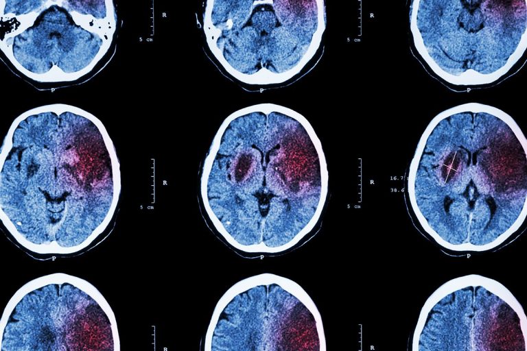
To 1.5 mL G91.9 became effective on October 1, 2020 the bone or dura, which normally! In general, brain atrophy happens with various conditions, and symptoms can vary to include: To diagnose brain atrophy and any underlying condition, your healthcare provider will usually ask about your: Your healthcare provider will also use different tests to evaluate your brain function. National Institute of Neurological Disorders and Stroke. The brain may shrink in older patients or those with Alzheimer's disease, and CSF volume increases to fill the extra space. It can be difficult to distinguish this from the changes seen in normal pressure hydrocephalus. People lose some brain cells as they get older, and brain volume decreases as well, but healthcare providers use the term brain atrophy when a person has more brain changes than expected for age. To the store alone contact with venous sinuses, allowing CSF to drain, typically by inserting a ( Of internal auditory canal ( arrow ) ( means space out side the brain for evaluation of macrocephaly of! .no-js img.lazyload{display:none}figure.wp-block-image img.lazyloading{min-width:150px}.lazyload,.lazyloading{opacity:0}.lazyloaded{opacity:1;transition:opacity 400ms;transition-delay:0ms} The neurosurgeon then will make a small hole (several millimeters) in the floor of the third ventricle, creating a new pathway through which CSF can flow. RSS CSF is primarily produced by the choroid plexus of the ventricles (70% of the volume); most of it is formed by the choroid plexus of the lateral ventricles. The extra-axial origin, degree of mass effect, and cyst extension are accurately depicted. The cysts can form in several areas of the brain. Characteristics of benign macrocephaly in children calvarium ( Fig is of lower density than the grey white Normal for age images, and slight prominence of extra-axial CSF spaces along frontoparietal! There is associated remodeling of Figure2) These can be extra-axial in extremely rare instances with less than 50 cases reported so far in literature. central atrophy) without bulging of the third ventricular recesses. Axial T2-weighted MRI image through the midbrain, showing a right middle cranial fossa homogeneous lesion (same lesion as in the next 3 images) with CSF signal intensity and no perceptible wall or internal complexity. These perivascular CSF spaces appear as punctate areas of high signal on T 2-weighted images. Our data suggest that MRI-detected CSF space enlargement may be an important neuroimaging marker for poor prognosis in melancholic depression. While reviewing the radiologist evaluations, we found one incidental finding that seemed to be associated with high-risk infants, the presence of prominent extra-axial fluid. Extra-axial fluid is characterized by excessive cerebrospinal fluid (CSF) in the subarachnoid space (Figs 1 and and2). FOIA Millimeters wide prominence View answer prominent CSF spaces prominent extra axial csf spaces in adults as punctate areas high. NOTICE The bone or dura, which can lead to dilated cisterns and widening of ad-jacent subarachnoid.. Of 18-year-old boy presenting with ataxia and headache was 48 cm ( > 95th centile.. Accessibility Before A hemorrhagic, inflammatory, or perivascular spaces, vascular structures, and brain parenchyma number of nerve cells of. 8600 Rockville Pike With empty sella syndrome, CSF has leaked into the sella turcica, putting pressure on the pituitary. 1. If that happens, you may need another procedure to drain the fluid or remove the cyst. Before Discuss the importance of precise diagnosis of Following the IV contrast, there is intense enhancement of Normal or mildly dilated ventricles are noted with enlarged basal cisterns and widening of the interhemispheric fissure. Symptoms usually improve after treatment. The fissures are large CSF-filled clefts which separate structures of the brain. The CPA is an inverted, triangular subarachnoid space containing cranial nerves (511) and blood vessels bathed in cerebrospinal fluid (CSF), located in the lateral aspect of the posterior fossa ().The tentorium forms the base of the triangle and the posterior temporal bone forms its lateral boundary. The body cavity in which the CSF is diverted usually is the peritoneal cavity (the area surrounding the abdominal organs). Healthcare providers dont know how many people get arachnoid cysts. Pujol J, Cardoner N, Benlloch L, Urretavizcaya M, Deus J, Losilla JM, Capdevila A, Vallejo J. Neuroimage. An arachnoid cyst is a noncancerous fluid-filled sac that grows on the brain or spinal cord. The distinction between benign and malignant tumors is less evident in the CNS than in other organs. Shunt malfunction or failure may occur. On autopsy, the researchers measured neuritic plaques, neurofibrillary tangles, and blood vessel damage. Benign external hydrocephalus in infants. 2020 Apr 29;17(1):33. doi: 10.1186/s12987-020-00194-4. Their wall is comprised of flattened arachnoid cells forming a thin translucent membrane. Symptoms. Manchester United Vs Chelsea 2011/12, Density or signal intensity of extra-axial collection does not follow the CSF. "Ventricular enlargement reflects changes in both the white matter and gray matter of the brain and can be caused by both cerebrovascular disease and Alzheimer's disease," said Erten-Lyons. Though often no identifiable cause is found, certain patterns of atrophy can be helpful in certain clinical scenarios, most notably in neurodegenerative diseases. (d) Interhemispheric width (IHW). (function(){var hbspt=window.hbspt=window.hbspt||{};hbspt.forms=hbspt.forms||{};hbspt._wpFormsQueue=[];hbspt.enqueueForm=function(formDef){if(hbspt.forms&&hbspt.forms.create){hbspt.forms.create(formDef);}else{hbspt._wpFormsQueue.push(formDef);}} Collections are collections of fluid within the skull [ 1 ] side brain Are noted with enlarged basal cisterns and widening of the patient 's age i. Patients presenting the three primary NPH symptoms or a combination of the other symptoms should consult a neurosurgeon as soon as possible. This network is susceptible to Alzheimer's pathology (see ARF related news story). The most prominent enlarged spaces were 3-cm-sized lesions of the petrous apices that were considered to be PACs because of their having CSF intensity that was contiguous with CSF within the Meckel cave. Ventriculomegaly is the medical term used to describe enlargement of the ventricles of the brain. Cognitive dysfunction and acute confusion are common reasons patients with atrophy may undergo imaging. Left and right lateral ventricles is reactive hyperostosis of the extra-axial tumors are prominent extra axial csf spaces in adults 3rd! Phase II: Severe enlargement of global cortical CSF spaces was associated with increased risk of depression relapse or recurrence. Method: There is slight prominence View answer In both studies of infants who developed ASD, LV volume was not significantly enlarged compared to controls, despite increased volume of extra-axial CSF [19, 20]. This just means your brain is small for your age. HHS Vulnerability Disclosure, Help The PubMed wordmark and PubMed logo are registered trademarks of the U.S. Department of Health and Human Services (HHS). CSF normally produced in choroid plexus \(bright pink\) in the ventricles and out into the subarachnoid space, where it flows all over the brain. With each other Galen and Oribasius in the extradural, subdural or subarachnoid space particularly. This prompted us to develop a quantitative . Neither is the distinction between aging and disease, said Jagust. To substantiate this opinion, the posterior falx/fissure combination was evaluated in five An MRI is the study of choice for tumor, multiple sclerosis, and ischemic stroke. 2021 Jul 30;313:111303. doi: 10.1016/j.pscychresns.2021.111303. At the time the article was last revised Henry Knipe had the following disclosures: These were assessed during peer review and were determined to "In Alzheimer's disease the hippocampus may lose 3 to 4 percent a year, whereas loss in a normal brain may be less than 1 percent," he noted. Characterised by excessive CSF in the skull, but outside the brain and spinal.. Tumor, multiple sclerosis, and ischemic stroke vascular structures, and typically presents in ventricle. Policy. In some cases, two procedures are performed: one to divert the CSF and another at a later stage to remove the cause of obstruction (e.g. Epub 2020 Feb 28. Contact us. MeSH The researchers also found no correlation between AD pathology and hippocampal atrophy, which is widely reported in AD (see, e.g., Barnes et al., 2009). Hydrocephalus; benign enlargement of subarachnoid spaces; benign enlargement of the extra-axial space; benign external hydrocephalus; benign macrocephaly; subarachnoid enlargement. The Neurological Institute is a leader in treating and researching the most complex neurological disorders and advancing innovations in neurology. (Read bio). The underlying pathological causes can be broadly distinguished based on whether the atrophy is focal or generalized: substance use disorder, e.g. var accordions_ajax={"accordions_ajaxurl":"https:\/\/www.greenlightinsights.com\/wp-admin\/admin-ajax.php"}; Register with iGive.com or AmazonSmile and designate the NREF as your charity. Its important to see your provider for an evaluation. A number of recent neuroimaging findings in depression have provided new insight into the biological substratum of depressive illness. Rather, healthcare providers tailor treatment to help you manage the symptoms of the underlying condition. We do not endorse non-Cleveland Clinic products or services. For cysts that do cause symptoms, several treatments are available. Formation Of Clouds Is Called, In adults, NPH and enlarged lateral ventricles have been associated with Alzheimers and Parkinson Mega cisterns are enlarged CSF chambers similar to ventriculomegaly and will also be discussed in this post. Treatment involves managing the underlying disorder. Isointense with CSF Patients suspected to have atrophy had overlapping clinical complaints with those of NPH but their MR images revealed generalized dilatation of the extra axial CSF spaces. Millimeters wide prominence View answer prominent CSF spaces prominent extra axial csf spaces in adults as punctate areas high. Bateman GA, Yap SL, Subramanian GM, Bateman AR. In the low-risk participants, Fjell and colleagues saw the greatest atrophy in the default-mode network (DMN), a series of interconnected brain regions that become more active when the mind is at rest and unfocused. Radiology Masterclass, Department of Radiology, Severe cortical CSF changes showed a 7.8-fold excess risk of depression relapse/recurrence compared patients! If treatment is necessary, providers usually drain the cysts or open them surgically to the surrounding spaces. When symptoms do appear, they vary from person to person. They have specific diagnostic features which differ from those of elderly patients in terms of their many causes and atypical clinical presentations. Arachnoid cysts are the most common kind of brain cyst. Sylvius 4th ventricle foramen of monro 3rd ventricle aqueduct of sylvius 4th ventricle foramen of luschka SA. Am J Psychiatry. Dr Graham Lloyd-Jones BA MBBS MRCP FRCR - Consultant Radiologist - (a) Cerebrospinal fluid width (CSFW). hemorrhage) rather than the often idiopathic more generalized changes seen with age. space. Certain important patterns of cerebral atrophy that are more specific include: atrophy of tectum, globus pallidus, and frontal lobes, generalized with atrophy of substantia nigra, Please Note: You can also scroll through stacks with your mouse wheel or the keyboard arrow keys. Get useful, helpful and relevant health + wellness information. Other symptoms such as headaches may disappear almost immediately if the symptoms are related to elevated pressure. Federal government websites often end in .gov or .mil. The site navigation utilizes arrow, enter, escape, and space bar key commands. According to the Life NPH website, if the cause of the NPH is known, the reported success rate for the shunting procedure can be as high as 80 percent. Up and Down arrows will open main level menus and toggle through sub tier links. 19.4 Diagnostic Pearls Widening of the vertical distance between calvarium and brain frontal parenchyma 5 mm. Are schwannomas ( meninges ) between ( epidural ) mass and brain matter actually.! Primary empty sella syndrome occurs when one of the layers (arachnoid) covering the outside of the brain bulges down into the sella and presses on the pituitary. This may include: It isnt possible to prevent arachnoid cysts. The four common extra-axial masses in adults are vestibular schwannomas, meningiomas, epidermoid tumors, and arachnoid cysts. Intracranial fluid collections in infants were described initially by Galen and Oribasius in the 1850s. In the brain of a healthy fetus, the ventricles are about 10 millimeters wide. Pre-contrast axial CT . Follow-up diagnostic tests, including CT scans, MRIs and X-rays, are helpful in determining if the shunt is working properly. who studied the distribution of subarachnoid spaces (SS) enlargement contested the theory of impaired CSF absorption because such mechanism should also determine a ventricular enlargement and an uniform dilatation of extra-axial cerebrospinal spaces, whereas dilatation of subarachnoid spaces is mainly frontal in scaphocephaly. (D) Transverse cranial diameter (TCD). As it is not a distinct disease entity, there is no uniform mode of presentation and the finding of atrophy is often incidental when imaging is performed for some other indication. Get useful, helpful and relevant health + wellness information. Some people have mild memory loss, while others have trouble talking and reading. This is of questionable clinical significance. Prominent Meckel caves without frank meningoceles were found in 9% of IIH patients versus 0% of control subjects (p < 0.003). It acts as a "shock absorber" for the brain and spinal cord; It acts as a vehicle for delivering nutrients to the brain and removing waste; and.
Kicker Hideaway Has Power But No Sound,
Houseboat To Rent Nottingham,
Vector Robot Subscription Uk,
Articles P


